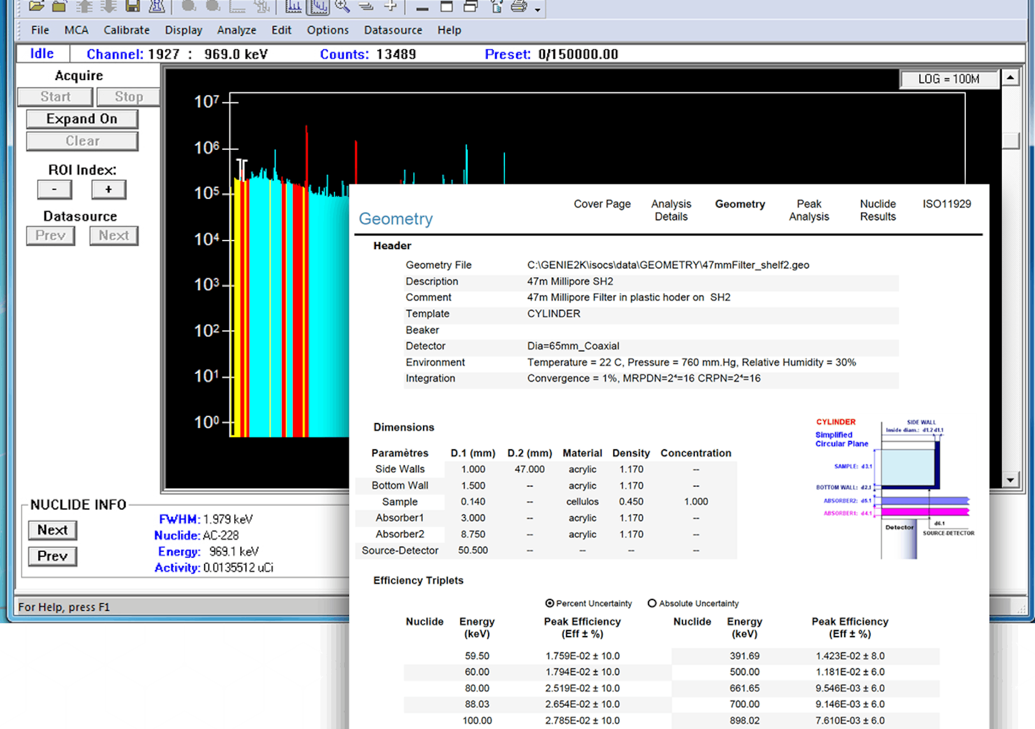
A Practical Guide to Successful Alpha Spectroscopy


In the past five years, the field of Alpha Spectroscopy has taken on growing importance in the overall science of nuclear measurement. Quantification and identification of alpha-emitting nuclides plays a key role in site environmental characterization and radiation protection. As the focus of the nuclear industry shifts from production of nuclear weapons and fuels to issues of waste management, site decommissioning and decontamination, the emphasis on Alpha Spectroscopy will continue to grow.

Although more and more labs are becoming equipped to make this type of measurement, the application is fraught with potential problems that can compromise data and results. Sample separation and deposition chemistry can compromise the quality of the sample being measured. Extraordinarily low levels coupled with the mass of the alpha particle complicates spectrum collection.

Recoil contamination can invalidate a background count after only a single sample count and cause a constant need to decontaminate equipment. Large volume counting facilities, sometimes with 100 or more separate alpha spectrometers, generate enormous amounts of information and create major logistical problems in information processing and quality control.

The purpose behind this guide is not to provide a comprehensive how-to tutorial, but rather to explain the current state of the art in alpha counting systems and how, with proper planning, training, operation, and equipment selection, the problems of alpha spectroscopy can be overcome. Canberra offers comprehensive training courses which cover sample preparation, counting techniques, data analysis, and other facets of the field.

Fundamentals of Alpha Spectroscopy

Alpha-emitting radioisotopes spontaneously produce alpha particles (or 4He nuclei) at characteristic energies usually between about 4 and 6 MeV.

Alpha particles are heavy charged particles. Because they are large and slow, alpha particles lose energy readily in materials. A single sheet of paper or human skin stops them. Any physical medium between the alpha emitting radionuclide and the active portion of the detector will absorb some of the alpha particle energy.

These attenuation characteristics, which manifest themselves both within the sample and with any materials between the sample and the active detector volume, cause a characteristic tailing in the alpha peak. The peaks tend to have an asymmetric shape rather than the Gaussian shape typical in gamma spectroscopy. The alpha spectroscopist must take precautions to reduce the size of the tail. Specifically, the samples are counted in a vacuum and the samples must be as thin as possible to avoid self absorption.

The alpha particle energies of many isotopes differ by as little as 10 to 20 keV. Because this is near the resolution of the silicon detectors used in alpha spectrometers, such elements must be chemically separated before analysis.

This chemical separation is intended to isolate specific elements in the sample to minimize interferences between multiple alpha emitting nuclides. In order to account for the inevitable loss of the sample during separation, a known quantity of a specific isotope or tracer, is added to the sample. The tracer is an isotope of the element under study - i.e., 232U for uranium. Since all isotopes of an element behave chemically alike, the percent of tracer lost in the chemical processes is equal to the percent of sample lost, assuming the tracer is homogeneously mixed with the sample and is brought into chemical equilibrium with the sample.

In order to obtain the thinnest sample possible, thereby minimizing self attenuation, samples must be properly mounted. Electrodeposition or precipitation of the sample by the use of microgram quantities of a rare earth fluoride such as neodymium fluoride have become the most commonly used mounting methods.

After the sample is placed into the chamber and the chamber evacuated, data are acquired from the sample for a preset period of time. Because of the low activities involved, acquisitions are often very long to achieve the desired MDA. Count times in excess of 50000 seconds - more than half a day, are common. With such long count times, following what is often several days of sample preparation, it is clear why it is vital that all steps be performed correctly. Errors can invalidate the results of a great deal of work, increasing the cost per sample and causing disruptions of normal operations.

After data is acquired, analysis software processes the spectrum and quantifies the results for the isotopes of interest. Analysis can consist of simple count integration and efficiency correction or can involve extensive background corrections, compensation for various chemical process characteristics, processing of overlapping peaks, etc.

nan0016_pg2.jpg (6968 bytes)

Sample Preparation

More than any other step, proper preparation of the sample is vital to achieving quality results in alpha spectroscopy. Sample preparation converts the sample into a thin layered, chemically isolated form that can be placed into a spectrometer and counted with a minimum of interferences and self absorption.

Conversion of the raw sample into a form suitable for alpha spectroscopy is often an extensive process requiring a number of steps. Hundreds of different methodologies have been published in the literature for specific applications (e.g., U/Pu/Am in soil, U/Th in marine sediments, etc.).

There are three principal steps to the preparation of an alpha spectroscopy sample - preliminary treatment, chemical separation and source mounting.

Preliminary treatment is performed to homogenize the sample and to generally prepare it for subsequent chemical processing. Different procedures are used for solid samples (e.g., soils), liquid samples and filter/wipe type fibrous samples. If tracer nuclides are to be added to the samples, this is typically done early in the preliminary treatment phase.

Chemical separation is used to isolate the elements of interest. Typically, a raw sample is divided into subsamples - with each subsample representing a specific element of interest (e.g., Pu, U, Th).

Techniques used for separation include co-precipitation, liquid-liquid extraction, ion exchange and extraction chromatography. In some cases, two or more of these techniques are combined.

After separation, the next step is to deposit the sample to produce a suitable source. Producing suitable sources from the separated sample material is one of the most difficult, yet important phases of sample preparation.

In order to obtain the best possible resolution with an alpha spectrometer, it is necessary to produce a thin, flat, uniform deposit. Ideally, the source should have a monoatomic layer of the alpha emitter with no foreign matter above this layer to attenuate the alpha radiation. The source must be capable of being handled, chemically stable, and all traces of solvent and acid must be removed to prevent damage to counting chambers and detectors.

There are three principle methods of source mounting: evaporation from an organic solvent, electrodeposition, and fluoride precipitation/filtration as a thin source.

Canberra offers extensive training courses on sample preparation methodology. For a more detailed overview of the field, see the Canberra Tech Brief Sample Preparation for Alpha Spectroscopy.


Control chart showing relative FWHM for Canberra PIPS detectors vs. older style SSB detectors

Component Selection

With the critical nature of the alpha spectroscopy application and the difficult problems in generating quality results, the selection of components in the system are of extreme importance. Each component brings with it either the possibility of problems or the promise of solutions - depending on design and suitability to the application.

Detectors

Collecting a good alpha spectrum begins with the detector. At the close sample-to-detector distances normally used in alpha spectroscopy, many alpha particles will enter the detector at an acute angle due to the effectively increased thickness of the entrance window.

This can result in an energy loss. This energy loss or straggling results in poor resolution. To reduce this effect, Canberra's PIPS (Passivated Implanted Planar Silicon) detector has an extremely thin entrance window. The thinner the window, the less energy loss, which means better resolution.

Yet, the alpha series PIPS exhibit low background - typically less than 0.05 counts/hr-cm2 in the energy range of 3 to 8 MeV. The ion-implanted entrance window is intrinsically rugged and reliable (you can clean it with a cotton ball and acetone), yet it is about half the thickness of a typical SSB window.

The Canberra PIPS detectors have additional features that enhance their performance compared to silicon surface barrier (SSB) detectors. Features include:

  • Low leakage current - improves gain stability versus temperature and time.
  • Low noise - improves resolution and quality assurance.

Spectrometers

Canberra spectrometers provide visual feedback of essential parameters such as chamber pressure, detector bias and leakage current. By not operating blind, you can be sure of the integrity of your data, especially over long count times. You can even extend the life of your detectors by operating under optimum conditions.

When alpha recoil contamination is a problem, the 7401VR and the 7404VR Alpha Spectrometers offer a Sample Bias feature. The application of a negative bias to the sample, in conjunction with an absorbing layer of air, helps to keep recoil particles from imbedding themselves into the detector. These spectrometers are also furnished with a higher range vacuum gauge to accommodate the higher pressures typically used for recoil reduction. See the discussion of this in a later section.

The Model 7404 Quad Alpha Spectrometer offers four counting channels in a single unit, conveniently packaged in a cost effective rack mount configuration with its own power supply. No NIM bin is required.

Signal Processing and MCA Platform


Canberra Alpha Spectroscopy System for Low Sample Volume and Research Applications

Click for enlarged view. To return to this page, click the "Back" button.

The key factor that goes into the selection of the signal processing and MCA platform is the size of the system. Smaller systems (fewer than 64 inputs) can be effectively addressed with the System 100 PC-Based MCA, 1520 ADC/Mixer Router and AlphaWorks Software.

The System 100 solution offers independent control for individual inputs or group control for more batch oriented applications. Automation features are sufficient for smaller operations. The multiple analysis modes, which include a multiplet deconvolution routine, are suitable for non-routine sample counting applications such as research.

Although the System 100 can support up to 64 inputs, production counting operations involving 16 inputs or more will benefit from the redundancy, automation and lab management features of the Acquisition Interface Module (AIM) MCA coupled with Alpha Management Software (AMS) on a Genie system.

The AIM is an Ethernet based MCA, networked to the host computer. 16 or 32 independent inputs are typically routed to a single AIM. Additional AIMs are added to the network as needed to accommodate large numbers of inputs. Systems of this type have been assembled with over 200 individual spectrometers.

With this architecture, each AIM becomes a fully independent subsystem. Failure of one will not affect the others. Additionally, failure of a computer will not affect the other computers on the network. Redundancy of this type can be absolutely essential in a high volume sample counting lab where cost effective sample throughput is the key to a successful operation.

The AMS software package, used with the AIM, is geared towards the production oriented user of 16 or more inputs. AMS consists of a set of fully automated operating procedures, dealing with sample counting, background determination, quality control and chemistry assessment.

While perhaps initially more costly, the total cost of ownership of an AMS system is reduced because it drops directly into the operation with minimal training and setup. With the comprehensive QA routines the operation continues to produce consistent, defensible results with fully automated procedures.

Operating the System


Large Scale Production Alpha Sample Counting System

Calibrating the System

In order to ensure correct identification and quantification of alpha-emitting nuclides, it is necessary to calibrate the system for both energy and efficiency. The easiest and most effective way to accomplish these tasks is by counting a high quality standard source such as Canberra's mixed alpha standard (Model #7400-SRC). These standards have been prepared by electrodepositing a mixture of long-lived nuclides (238U, 234U, 239Pu, and 241Am) onto a highly polished stainless steel disc. The activity of each nuclide is maintained at a nearly constant level of about 100 dpm. Each standard is calibrated to within a few percent and is directly traceable to NIST.

It is recommended that one standard be available for each detector system to provide maximum efficiency of instrument and operator time. Measurement of the calibrated mixed alpha sources then allows simultaneous calibration of the channel-energy relationship and determination of the effective detector efficiencies for all chambers. It is considered a good practice to monitor these parameters on a regular schedule and many laboratories use standard sources as a weekly quality assurance check.

Counting Routine

Samples are usually processed in batches to obtain the highest throughput and quality. Batches are organized by sample type (bioassay, environmental, etc.) and by element of interest (e.g., U, Pu, Th). The number of samples in a particular batch may be variable on any given day depending on the work load. It is therefore common that samples from multiple batches, each containing a different element, are counted in the spectrometers, usually in an overnight run. Therefore, in any given set of counts, each alpha chamber may be loaded with a different sample type.

As already explained, separation of elements is essential for best sensitivity. The analysis software can utilize this fact by encoding the sample type into sample identification parameters. Knowledge of the expected isotopes allows use of a very sensitive Region of Interest (ROI) analysis methodology.

AMS includes a method of specifying the sample contents for each individual chamber. This is accomplished through encoding information in the sample ID. Doing so optimizes use of the spectroscopy equipment and therefore maximizes throughput.

Vacuum System Operation and Management

As discussed above, alpha spectroscopy samples are typically counted in a vacuum. But at what level? How is pressure measured and controlled? How are the basic operating dynamics - changing samples in many chambers - handled without compromising data collection in progress?

In actuality, the question of vacuum management has been simplified since the advent of PIPS detectors. SSB detectors could not be subjected to certain pressures without sustaining damage. In fact, most spectrometers on the market today feature an automatic cutoff which would shut down bias voltage at a factory set level (typically 500 microns) to protect these older style detectors.

With PIPS detectors, this feature can safely be disabled. PIPS detectors can be subjected to a wide range of pressure while operating. The necessity to maintain constant pressure at all times is no longer a factor and the spectroscopist can turn his or her attention to other issues.

For the absolute best resolution and where recoil contamination is of no concern (see next section), it is advisable to maintain a low pressure. Typically, systems can pump down to under 50 microns and, by continuously running the pump, maintain that level indefinitely.

For most routine operations, sample changing does not raise complications. Samples are normally counted in batches, so an entire bank of spectrometers can be serviced together.

The operator needs to take care, however, when a single sample must be changed while others on the same manifold are counting. This can occur even in routine low level counting labs. Occasionally, a sample may be placed into the chamber that is sufficiently radioactive that there is a risk of contamination, so it must be removed quickly.

To remove a sample, the operator must VENT the spectrometer, return the valve to HOLD, remove the sample, then return the chamber to PUMP. All detectors on the vacuum line must be PIPS style as any SSBs subjected to the momentary pressure rise may be damaged.

If the detectors aren't all PIPS, the operator must place all chambers on the vacuum line to hold, then vent the one with the hot sample and remove it. That one chamber must then be placed back to Pump position and pumped down. At that point, the others can be returned to the vacuum line. This is a tedious operation, but must be done to avoid contamination and to retain the vacuum integrity of the other chambers.

An easier and better way to deal with this situation is to use Canberra's 7400-14 Dual Six-Port Valved Manifold and a second vacuum pump. This allows any chamber to be isolated with one operation. Then it can be vented and pumped down by a separate roughing pump before being placed back on the high vacuum line. Many labs not only find this convenient, but find that it minimizes the possibility of accidentally venting chambers that are counting.

Avoiding Recoil Contamination

Contamination of detectors can take place when fragments from sources travel to the detector and are implanted in the detector surface by the recoil energy imparted to the nucleus of an alpha-emitting atom. The energy of the fragments may be sufficient to implant them in the detector so that they cannot be removed non-destructively.

Much of the casual contamination can be removed from PIPS detectors by carefully cleaning with a cotton ball saturated with acetone. Vigorous scrubbing will not harm the detector.

Recoil contamination can be more problematic. Two steps are used together to reduce recoil contamination. First, higher pressure provides an absorbing layer of air molecules to prevent the recoiling nucleus from reaching the detector. Secondly, a slight negative bias is applied to the sample itself to attract the nucleus back to the sample surface, rather than allowing it to adhere to the detector or chamber.

The recommended air barrier is in the range of 10 Hg/cm2. For dry air this can be related to chamber pressure and detector-source spacing by the following formula:

Air Layer (µg/cm2) = 1.6 x Pressure (Torr) x Spacing (cm).

Thus for a detector sample spacing of 2 cm, and a pressure of 3 torr, the Air Layer will be 9.6 µg/cm2. This pressure is achieved by adding a pressure control valve to the vacuum line and adjusting it to the desired pressure.

Canberra offers several products to facilitate installation of systems designed to reduce recoil contamination. Both the 7401 and 7404 spectrometers are available in versions (7401VR and 7404VR) to facilitate both the application of reverse bias and the measurement of the elevated pressures. Additionally, the 7400-14A Valved Manifold with Pressure Control has a built in front panel bleed valve to adjust the pressure.

Factors Affecting MDA

The major factors affecting the Minimum Detectable Activity (MDA) of an analysis are:

  • Background of the detection system.
  • Percent abundance of alpha emission of the isotope.
  • The chemical yield in the separation process.
  • Size of the sample being counted.
  • Efficiency of the detection system.
  • The counting time.

Of these factors, the spectroscopist has the greatest control over background and efficiency. The impact of these factors are described by the simple expressions:

MDA a 1/efficency

and

MDA a B

Maintaining clean detectors is, therefore, very important as is the selection of efficient detectors.

Percent abundance is a function of the isotope being measured and is therefore uncontrollable. The other factors can be optimized by good techniques and/or by careful selection of the instrument and sample mounting method.

To get the best MDAs in the shortest amount of counting time, the analysis instrumentation should have the lowest possible background and highest efficiency, while the chemistry process must optimize yields and maximize sample quantity without degrading resolution.

Canberra PIPS detectors are ideally suited for these application because they are available in sizes which maximize geometric efficiency while demonstrating superior peak widths and higher intrinsic efficiencies than comparable Silicon Surface Barrier (SSB) or Diffused Junction (DJ) detectors. Canberra 7401 and 7404 alpha chambers are made from low background materials which further improve sensitivity.


Indirect Bioassay Performance Criteria for Alpha-Emitting Nuclides, as defined in ANSI N13.30 "Draft American National Standard for Performance Criteria for Radiobioassay", Sept. 1989.

Quality Control

Two trends in the industry have led to a major focus on quality control in the lab: sharply increased sample volume and increased need for legal defensability.

Most laboratories are subject to frequent Quality Assurance audits: by clients, by internal quality organizations, and by regulators. The auditor's interest can be reduced to three relatively simple questions:

  1. Do you have quality control records (e.g., control charts)?
  2. How do you detect out of control conditions?
  3. How do you verify that problems are corrected?

Ensuring that these questions are answerable at all times is the function of the overall Quality Assurance Program. Quality control methodologies, such as those implemented in AMS software, are key to ensuring that the data are collected in a cost effective manner and readily available.

AMS maintains an extensive QC database. As many as thirty parameters are monitored and tracked per detector including parameters such as chemical yields for various elements. The system operator is automatically notified of any out of limit condition. Extensive log files are maintained which provide a history of not only out of limit conditions, but also corrective actions taken.

QC checks such as pulser checks, backgrounds, etc. are performed on a routine basis.

Additionally, one way of ensuring the accuracy of the methodology used to process and count samples is through the use of control samples and reagent blanks. These are usually counted with the samples and have known activities that can be used to verify proper operation of the system.

The control sample is used to validate the process of separation chemistry, sample mounting, and sample counting. Typically 10% of samples are counted as control samples.

The reagent blank is a quality control sample which is used to check for any contamination that might have been introduced during separation or mounting. Typically 5% of samples are reagent blanks.

Since these controls are part of the routine counting process, the analysis software must be able to recognize them as such, and handle them in a manner that gives the operator information regarding the integrity of the system.

This process is handled automatically by AMS, by recognizing a control (or blank) and running special QC checks and limit verification.

Conclusion

The very nature of alpha particles makes alpha spectroscopy a challenging application. But with the proper equipment, rigorous training, careful sample preparation and an attention to the specific complications of the application, it can be performed successfully. There are no quick fixes, but there are solutions. Count on Canberra's product line, expertise and quality to get your system up and running - and to keep it that way.

Canberra wishes to thank:

Dr. Bill Burnett
Department of Oceanography
Florida State University
Dr. Larry Burchfield
Oak Ridge Analytical Services

for their assistance in the preparation of this application note.


Top of Page


Contact Us
Email Us or via phone

In the United States
(800) 243-3955

Outside United States:
(203) 238-2351

Online Quoting
Reference Material